The laboratory
GHK-Cu experiments in cell movement and repair
Repair has verbs: move, pull, make, remove. I can follow each action through an experiment; a collection of proposed benefits gives those actions too little room.
Movement and contraction
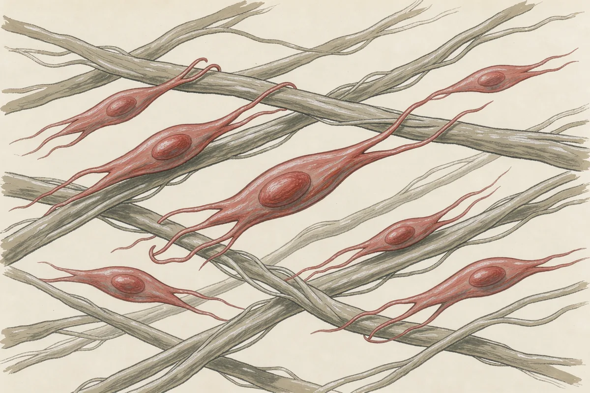
The aged-mouse fibroblast study used free GHK. Cells migrated more readily and contracted collagen gels more strongly in a dose-dependent response. Markers p21 and p53 decreased, while p63 and PCNA were restored. The researchers connected those changes with an altered aged-cell state and proposed integrin-beta1 signalling as a mechanism.
A collagen gel supplies a material the cells can act on. That makes this assay unusually satisfying to picture: a living cell changes its surroundings. Still, contraction is the measured task. Calling the result whole-body age reversal would give the experiment a job far larger than the one it performed.
The next experiment could connect those cellular behaviours with the strength and function of repaired human tissue. For now, the useful finding sits in cultured cells from aged mice.
Collagen output from fibroblasts
In Maquart and colleagues’ 1988 experiment, GHK-Cu stimulated collagen synthesis in human fibroblast cultures. The cell number stayed unchanged. Production began to rise between 10^-12 and 10^-11 M and reached its maximum at 10^-9 M in that experiment; these are concentrations in culture medium.
That unchanged cell count matters. A bigger population could have produced more collagen without each cell doing anything differently. Here the result pointed to changed output from the existing cells.
The attraction of a collagen result is obvious. The harder question is how that material is organised and used. A cell’s production figure and a healed tissue’s performance measure different parts of the work.
Matrix turnover
The MMP-2 study in fibroblast cultures adds the removal side of repair. GHK-Cu increased expression of that enzyme and its messenger RNA, with concurrent increases in the inhibitors TIMP-1 and TIMP-2. Free GHK failed to reproduce the effect.
The extracellular matrix is the supporting material outside cells. Enzymes can cut it back while other processes rebuild it. A slogan about “boosting collagen” leaves out the coordinated turnover. I prefer the busy account, enzymes and restraints included.
Rat wound dressings
A 2005 rat wound experiment incorporated biotinylated GHK into a collagenous matrix and found accelerated dermal healing. Biotinylation is a modification of the peptide; the dressing was a prepared material as well as a way to hold it at the wound.
That setting gives the research a practical shape. I credit the prepared matrix alongside the modified peptide; ordinary GHK-Cu remains a separate preparation. Human scar-fading and scar-size outcomes remain unestablished by this animal work.
Gene-expression tallies
The 2018 gene-data review described broad shifts in GHK-related gene activity using cell data and Connectivity Map analysis. At the 50%-or-greater change threshold, 59% of affected genes increased expression and 41% decreased. In the protein-cleanup group, 41 genes increased and 1 decreased.
The familiar estimate of about 4,000 affected genes extends beyond the roughly 2,100 counted at that threshold. These tallies concern gene activity. They supply hypotheses about repair pathways, with protein-level and living-tissue confirmation still needed.
Give me a cell doing something observable. Gene data can help explain that behaviour; my enthusiasm for counting altered genes runs out well before the largest tally. Much of the foundational work also comes from Pickart and colleagues, so confirmation by other teams carries particular weight.
Inflamed intestinal tissue
A 2025 colitis experiment in mice found lower disease activity, preserved colon length and increased tight-junction proteins after GHK-Cu. Those proteins help maintain the joins between neighbouring cells. The study also reported reduced inflammatory signals through the SIRT1/STAT3 pathway.
This is another kind of repair task: maintaining a lining. It broadens the experimental questions, while evidence for treating human bowel disease remains absent. The human skin findings are a separate body of work, with different preparations and outcomes.